What to expect from a kidney biopsy for C3 glomerulopathy
 Reviewed by
A kidney biopsy is a standard test used to establish a definitive C3 glomerulopathy (C3G) diagnosis.
During the procedure, a doctor inserts a needle into the kidneys to collect a small tissue sample, typically using ultrasound for guidance. The physical sample extracted during the process is required to confirm a C3G diagnosis and make decisions on subsequent treatment options.
Learning what to expect from a kidney biopsy for C3G can ensure you are fully prepared for the journey ahead.
Why a kidney biopsy is essential for C3G
Although standard urine and blood screenings are helpful for identifying general renal issues, such as the presence of protein in urine (proteinuria), they are unable to verify the presence of C3 deposits alone. A kidney biopsy is the only test that conclusively shows the characteristics of C3G.
The condition results from an overactive part of the immune system called the complement system, which consists of a collection of proteins in the blood that help the immune system. As a result of this overactivity, deposits of C3 collect in the kidney and damage the tiny filtering units called glomeruli.
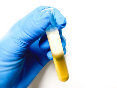
A doctor will examine the tissue extracted during a kidney biopsy under a microscope to see if there are abnormal levels of C3 protein deposits, without the presence of other proteins. This distinct pattern helps to identify C3G and rule out other kidney diseases.
Preparing for a biopsy procedure
The following steps will likely be taken to prepare for a C3G kidney biopsy:
- Complete preliminary lab work: The medical team will typically collect blood and urine samples before the procedure to confirm there are no active infections or another condition.
- Pause blood-thinning agents: Patients will be directed to stop using over-the-counter pain medications that thin the blood, such as ibuprofen, naproxen, and aspirin, for a full two weeks leading up to the appointment. These substances alter how the blood clots and increase bleeding risks.
- Stop specific supplements: For the same bleeding risk reasons, the care team will likely instruct the patient to stop taking certain dietary supplements, such as fish oil.
- Fast before the appointment: People may be asked to avoid all food and beverages for eight hours prior to the extraction.
What happens during the kidney biopsy
A kidney biopsy requires specific clinical steps to safely collect a small sample. It is most commonly done percutaneously, in which a small needle is inserted through the skin into the kidney to extract the sample. The procedure typically requires around one hour to complete.
Percutaneous kidney biopsy procedure
Someone undergoing a standard percutaneous kidney biopsy procedure can expect the following as part of the process:
- Sedation: Depending on the specific situation, individuals may undergo the procedure awake with local anesthesia, or be completely asleep under general anesthesia.
- Position: The patient will generally rest face down, supported by a firm pillow or sandbag placed beneath the rib cage. However, if the biopsy is being performed on a transplanted kidney, they will need to lie on their back instead.
- Procedure: The doctor inserts a special needle through the skin and guides it into the renal tissue, typically using ultrasound or X-ray images to locate the exact extraction site. They may ask the person to hold their breath during insertion. The patient may feel pressure as the needle is passed, but there should not be sharp pain. An audible “pop” or click when the needle takes the small piece of kidney tissue is completely normal.
After the procedure, stitches are not required. Instead, a doctor will apply a bandage to the needle insertion site.
Alternative surgical methods
If an individual has a history of bleeding problems and cannot safely undergo the standard percutaneous method, a surgeon may perform an open biopsy operation to actually look at the kidney and collect the sample.
In other instances, a doctor may perform a laparoscopic kidney biopsy by making two small incisions in the back and inserting specialized tools to extract the tissue while the patient is under general anesthesia.
Transjugular biopsy is another less frequently used method, in which the physician guides a catheter and needle through a vein in the neck directly into the kidney.
Post-biopsy recovery and immediate care
Following the extraction, the body requires strict rest to help the puncture site heal. Te ensure a safe kidney biopsy recovery, patients must adhere to these guidelines:
- Immediate clinical rest: Remaining still in bed for several hours in the hospital promotes healing at the insertion site and lowers the risk of internal bleeding. Patients can eat and drink during this time.
- Vitals monitoring: During rest, medical staff will frequently check blood pressure, pulse, and blood tests to monitor for any internal bleeding.
- Overnight observation: The medical team may require an overnight stay to watch for complications, though individuals should be permitted to leave the following day if their vitals are stable.
- Physical activity restrictions: People recovering from a kidney biopsy must avoid strenuous exercise, heavy lifting, sexual intercourse, and contact sports for at least several days
In the event of an open biopsy procedure, a healthcare professional may provide specific recovery instructions.
Individuals should seek emergency medical help right away if they experience any of these red-flag symptoms after a biopsy:
- Inability to urinate
- Urge to pass urine very often
- Burning sensation while urinating
- Urine that is dark red or brown
- Urine that has blood clots
- Increased pain at the biopsy site
- Skin color changes, warmth, or oozing from the biopsy site
- Fever
- Dizziness
- Feeling faint
Understanding biopsy results and next steps
After the extraction is complete, the tissue sample is sent to a pathologist for analysis. It typically takes three to five days for the pathology lab to provide complete results, though partial reports are sometimes available within 24 hours.
Pathologists use immunofluorescence to measure the degree and location of C3 protein staining within the kidney. An official C3G diagnosis is confirmed when the biopsy shows a large amount of C3 deposits inside the glomeruli, without the presence of other proteins called immunoglobulins.
Pathologists use an electron microscope to distinguish between the two primary forms of the disease, dense deposit disease and C3 glomerulonephritis, because they appear visually distinct under high magnification.
C3G News is strictly a news and information website about the disease. It does not provide medical advice, diagnosis, or treatment. This content is not intended to be a substitute for professional medical advice, diagnosis, or treatment. Always seek the advice of your physician or other qualified health provider with any questions you may have regarding a medical condition. Never disregard professional medical advice or delay in seeking it because of something you have read on this website.